Saturday, October 31, 2015

UNIZIK MED LAB LECTURER WINS UNESCO PRIZE

A Medical laboratory science lecturer in NAU, Dr. Rebecca Chinyelu Chukwuanukwu has emerged the first prize-winner of this year's UNESCO-Merck Africa Research Summit (UNESCO-MARS) held in Geneva, Switzerland recently.

Chukwuanukwu whose area of specialization is Immunology, beat 99 other scientists/researchers across Africa who attended the summit. The annual summit aims to bring together young top researchers across the continent to discuss the generation, sharing and dissemination of research data and to draw a roadmap for Africa's development as an international hub for research excellence and scientific innovation.

This year's summit focused on building research capacity in health and life sciences with special attention on Ebola and emergent infectious diseases. Chukwuanukwu's work was on: Modulation of the Immune Response to Mycobacterium Tuberculosis during HIV/Malaria Co-infection. The first prize comes with a prestigious six months fellowship in Merck research and development hub.

Source: Daily sun, Fri. Oct. 30, 2015

UNIZIK MED LAB STUDENT WINS MISS ANAMBRA 2015


UNIZIK STUDENT WINS MISS ANAMBRA 2015
Miss Blessing Chidinma Okeke, a Med Lab 200 level (entering 200) student of Nnamdi Azikiwe University has won the crown as the new Miss Anambra. She took over from another Zikite, Miss Chizoba Ejike from the Department of Accountancy. 
The Directorate of Public Relations of the College of Health Sciences Student Association Government on behalf of the government and the entire students of the college wish to say a big congratulations to her. 


Queen Chidinma Okeke, representing Orumba North, has been crowned the winner of the Miss Anambra 2015 pageant, organised by Anambra Broadcasting Service and the State government.

The beauty queen was crowned at the grande finale of the pageant, held at Marble Arch Hotels, Awka.
Correspondents Ejike Abana and Bright Nnachi, report, that In what may be best described as a night of glamour and display of beauty and brains, the top twenty one finalists took to the challenge floor as they fought to meet stringent requirements as set by the judges.

The girls made their first judging appearance in traditional attire, and their second judging appearance in casual outfit.
The third judging appearance was made in evening dresses, where top ten girls were chosen by the judges.
The end of a question and answer session saw the elimination of five girls, with Miss Njikoka, Orumba North, Ayamelum, Anaocha and Ihiala making the top five list.
Speaking, the Managing Director and Chief Executive Officer of Anambra Broadcasting Service, Mr Uche Nworah expressed appreciation that the pageant, which has the objective to use the platform to empower women, while also celebrating the elegance, beauty and grace of the Anambra woman, finally became successful, noting that the pageant in its second season, ranks as one of the biggest state pageants in Nigeria.

Mr Nworah thanked the co sponsors of the pageant for making the day fruitful and eventful, including the State government among others.

Speaking shortly after the event,  Miss Nigeria 2003/2004 and creative director of the Miss Anambra Pageant, Miss Nwando Okwuosa, in expression of happiness at the success of the event, advised the current Miss Anambra queen to make the best of the opportunity, be a good role model to people, especially the younger ones.

Earlier in a farewell speech, the erstwhile Miss Independence Anambra 2014 and the current Miss ECOWAS 2015, Queen Chizoba Ejike, stressed that the office of a beauty queen is not for women of easy virtues, urging the queen to use her office to reach out to the voiceless and less privileged.

Miss Anambra 2015, Queen Chidinma Okeke in excitement appreciated the ABS boss and other people that helped get her where she is, saying that the vision of introducing light into the lives of young women, through organising the pageant which wouldn't have been possible without the successes recorded by Governor Willie Obiano is a big plus.
Miss Okeke, who is from Ogboji in Orumba South, Local Government and a 200 level Medical  Laboratory student of Nnamdi Azikiwe University,promised to infuse her best to humanity, contribute immensely to the promotion of the organizers of the event, the ABS, and to propagate the rich cultural heritage of Anambra state and Nigeria.
She also pledged to be good example, role model to other Nigerian girls and a good ambassador of the state, assuring of full commitment, diligence and hard work.
The event also featured the presentation of awards to Miss Awka North, as the most photogenic contestant, Best Dinner Gown to Anyamelum, Best Model to Anambra East, Miss Amity to Ihiala, Best Traditional Costume to Ekwusigo, Most Intelligent to Dunukofia and People’s Choice  Award to Onitsha South Local Government Area.
Miss Anambra will represent Nigeria in Miss Intercontinental in Germany, come December.
The event attracted top government functionaries, including Commissioners, LGA Chairmen, Commissioner of Police, Mr Hassan Karma, a veteran Hollywood actor, Chief Pete Edochie, among others.
Comedy, music performances from notable artists such as Slow Dogg, Obiwon, Djinee, Twocantalk, Mc Parrot Mouth, dance performances by the girls, crowning of Miss Anambra 2015, among others formed the high points of the event.
Queen Chidinma Okeke was later presented with the Keys of a brand new KIA RIO car, by Dana motors ltd, a cheque of one million naira, wardrobe allowance, flight business class ticket by Air Peace ltd to travel anywhere in Nigeria for one year, among others.
Source: ABS


Saturday, October 24, 2015

TO THE FRESHERS AT OAKLAND


You have made it to the College Of Health Sciences, Nnamdi Azikiwe University, Nnewi Campus. You have struggled in your sojourn at the Awka Campus from MPH to Science Village and any other like the utility building, then back to MPH. Having met the minimum requirement for crossing over, you have earned a place in the campus.
The Nnewi campus of NAU is divided into the Okofia campus (which we refer to informaly as Oaklad), and the NAU Teaching Hospital located at the central part of Nnewi town (and you can guess why). Every fresher schools in Okofia so that's where our focus is.
Unlike the main campus, the college whose Provost is Prof. A.M.E. Nwofor is a small student community and with time you'll be able to recognise at least 65 percent of the students. So, on your arrival, apart from the little adjustments you'll have to make in adapting to the dress code and the JJC attitude you'll definitely display here and there either individually or as a group which will mmediately give you away, you can still be spotted as a fresher just by the unfamiliarity of your face.
Every fresher is a student of the Faculty of Basic Medical Sciences with respect to academic workload for the next one year. You'll all be studying Human Anatomy, Physiogy, and Biochmistry under the respective departments with Prof. C.S. Ufearo as Dean. You may also have one or two other courses from any other department especially your own department. Except in 2013 due to the ASUU strike, the department of Biochemistry usually sets an incourse assesment quiz before in at least one of their courses before the Xmas break. It might not be different this time.
You have worked hard at Awka, you'll have to work hard here too if not harder. The college library is always available at appropriate hours with fairly conducive atmosphere for study. You might not have attnded any lectures at Awka, but a minimum attendance of 75 percent to lectuures is required here. You may not have played or had any fun at Awka or you may have been involved in other's activities, but over here, get ready to join in the various actiities that would be going on from the first day of school some of which would affect you directly. In being a balanced student, you first must be a student, then strike a balance.
Accomodation in Okofia is not a big problem - you can not run out of choices. There is no school hostel for boys and the girls' hostel is very limited. That not withstanding, if you have not yet gotten your own place by now, you are wrong in my own opinion. Inasmuch as the older students have come to help you out with accomodation in one little way or the other, you are not to be blamed entirely. However, it's not too late to get one. The point is that you should try and settle down before the session begins so you can have no reason to be unsettled afterwards. Do well to identify with any Xtain body of your choice, they'll be of good help to you at this time.
Finally, men and brethren, ad all those that fear the Lord, this is the place that would likely mould and shape you for most of the rest of your life towards being a learned and disciplined individual and a responsible citizen. Here, you learn and unlearn, you develop and mature, you experience and experiment, you live and let live.
GOODLUCK TO YOU

From yours truly,
Onyenemezu Kelechi
400L Medicine and Surgery
*Don't fail to share to as many media outlets that all may be informed*

Friday, October 23, 2015

CRIME: 2 UNIZIK students hack into Vice President's account, steal N8Millon

Policemen attached to the Special Anti-Robbery Squad (SARS) Ikeja are investigating an alleged theft of N8million from Vice Pres ident Yemi Osinbajo’s bank account.

    It was gathered that two undergraduate students of the Nnamdi Azikiwe University Awka identi fied as Ebuka Orumba and Patrick Nwafor allegedly hacked into the account of Simmons Cooper Partner, a law firm said to belong to Vice President Osinbajo and allegedly stole eight million naira (N8miilion) from it.

    According to investiga tion, the undergraduates Nwafor is a final year 500level student of Envi ronmental Science while Orumba is a final year Computer Engineering student.

    It was gathered that the students used fraudulent means to hack into the email of the Account man ager of the VP’s law firm, one Mr. Tunde Irukera, and they allegedly sent a mail to UBA PLC branch to one Obuba Fidelia on July 2nd instructing the bank to transfer eight mil lion naira from Simmions Cooper Bank account to three different bank ac counts they opened for the fraud.


    It was further gathered that the undergraduates allegedly connived with a tricycle rider one Artor Kalu to open a bank ac count with Zenith bank ac­count number 2086943174 where three million naira (N3,000,000) was trans ferred to with the false name Engineer Asukwo Okoro.

    The remaining amounts were said to have been transferred to two other unnamed bank accounts.
    Luck however ran out on the undergraduates when their alleged gang member, Mr. Kalu went to the Zenith bank account at Mushin to withdraw the N3,000,000 but was nabbed by detectives from SARS following a tip off from the bank.

    The police subsequent ly arrested Kalu and fol lowing his statement, the police arrested Orumba but Nwafor escaped.

    Following the investiga tion, the police discovered that Kalu with the help of others got a fake Driver’s license which he used to open the Zenith Bank ac count.

    The police has since charged Orumba and Kalu to court on a 5 count charge on conspiracy, stealing and fraud while efforts is on to arrest Nwafor whom the others claimed to be the master mind of the fraud.


https://www.today.ng/news/27723/hackers-steal-n8m-from-osinbajos-bank-account

UNIZIK releases Direct Entry screening test results

This is to inform all candidates who participated in today's Direct Entry screening test that the results have been released. You can view your score on my.unizik.edu.ng/postutme/putmeresult_engine.php


Dr. Emmanuel Ojukwu

Director of Information and Public Relations


Thursday, October 22, 2015

Why UNIZIK shifted resumption date revealed

The Director of Public relations and Information, Dr. Emmanuel Ojukwu has revealed why the university management shifted the university calender. In a reply on his Facebook page, the Dr. Ojukwu wrote: "The Govt policy of TSA which mopped all university money into the central bank is the reason. As at now the university does not have access to its money. It is needless bringing students back when the university cannot offer basic services. We expect that by this new date we would have made adequate arrangement for services. This definitely is not the fault of the university. Many other Federal agencies are affected".
The new calendar released by the university management is below
NNAMDI AZIKIWE UNIVERSITY, AWKA
ACADEMIC CALENDAR FOR THE 2015/2016 SESSION (REGULAR STUDENTS)

The Vice-Chancellor has, on behalf of Senate, approved the Calendar for 2015/2016 academic session for regular students as follows:

FIRST SEMESTER
1. Monday November 9, 2015 New Students Return and Continue Payment of Fees and Registration
2. Monday November 23, 2015 Old Students Return
3. Monday November 23, 2015 Lectures Commence for all Students
4. Wednesday November 25, 2015 Senate Meeting
5. Monday November 30 – Wednesday December 2, 2015 Orientation of new students
6. Tuesday December 15, 2015 University Wide Quiz
7. Tuesday December 22, 2015 – Friday January 4, 2016 Christmas Break
8. Tuesday January 5, 2016 Lectures Continue
9. Wednesday January 27, 2016 Senate Meeting
10. Friday January 8, 2016 Matriculation
11. Monday February 21 – Friday February 25, 2016 Revision Week
12. Tuesday March 1, 2016 First Semester Examinations Commence
13. Wednesday March 2, 2016 Committee of Deans Meeting
14. Wednesday March 16, 2016 Senate Meeting
15 Wednesday March 23, 2016 First Semester Examinations End

SECOND SEMESTER
1. Friday March 25 – Tuesday March 29, 2016 Easter Break
2. Wednesday March 30, 2016 Lectures Commence after Easter Break
3. Wednesday April 6, 2016 Committee of Deans Meeting
4. Wednesday April 27, 2016 Senate Meeting
5. Thursday May 5, 2016 University Wide Quiz
6. Wednesday May 25, 2016 Senate Meeting
7. Monday June 13 – Friday June 17, 2016 Revision
8. Wednesday June 15, 2016 Committee of Deans Meeting
9. Monday June 20, 2016 Second Semester Examinations Commence
10. Wednesday June 22, 2016 Senate Meeting
11. Friday July 8, 2016 Second Semester Examinations End
12. Monday July 11 – Wednesday July 13, 2016 Project Defence
13. Monday July 18, 2016 Submission of Degree Results
14. Wednesday July 20, 2016 End of Second Semester/Senate Meeting
15. Thursday July 21, 2016 Long Vacation Commences

This supersedes the earlier calendar dated 18th September, 2015.

Dr. I. H. Isidienu, FCAI
Ag. REGISTRAR

NNAMDI AZIKIWE UNIVERSITY, AWKA ACADEMIC CALENDAR FOR THE 2015/2016 SESSION (REGULAR STUDENTS)


The Vice-Chancellor has, on behalf of Senate, approved the Calendar for 2015/2016 academic session for regular students as follows:

FIRST SEMESTER
1. Monday November 9, 2015 New Students Return and Continue Payment of Fees and Registration
2. Monday November 23, 2015 Old Students Return
3. Monday November 23, 2015 Lectures Commence for all Students
4. Wednesday November 25, 2015 Senate Meeting
5. Monday November 30 – Wednesday December 2, 2015 Orientation of new students
6. Tuesday December 15, 2015 University Wide Quiz
7. Tuesday December 22, 2015 – Friday January 4, 2016 Christmas Break
8. Tuesday January 5, 2016 Lectures Continue
9. Wednesday January 27, 2016 Senate Meeting
10. Friday January 8, 2016 Matriculation
11. Monday February 21 – Friday February 25, 2016 Revision Week
12. Tuesday March 1, 2016 First Semester Examinations Commence
13. Wednesday March 2, 2016 Committee of Deans Meeting
14. Wednesday March 16, 2016 Senate Meeting
15 Wednesday March 23, 2016 First Semester Examinations End

SECOND SEMESTER
1. Friday March 25 – Tuesday March 29, 2016 Easter Break
2. Wednesday March 30, 2016 Lectures Commence after Easter Break
3. Wednesday April 6, 2016 Committee of Deans Meeting
4. Wednesday April 27, 2016 Senate Meeting
5. Thursday May 5, 2016 University Wide Quiz
6. Wednesday May 25, 2016 Senate Meeting
7. Monday June 13 – Friday June 17, 2016 Revision
8. Wednesday June 15, 2016 Committee of Deans Meeting
9. Monday June 20, 2016 Second Semester Examinations Commence
10. Wednesday June 22, 2016 Senate Meeting
11. Friday July 8, 2016 Second Semester Examinations End
12. Monday July 11 – Wednesday July 13, 2016 Project Defence
13. Monday July 18, 2016 Submission of Degree Results
14. Wednesday July 20, 2016 End of Second Semester/Senate Meeting
15. Thursday July 21, 2016 Long Vacation Commences

This supersedes the earlier calendar dated 18th September, 2015.

Dr. I. H. Isidienu, FCAI
Ag. REGISTRAR


Wednesday, October 21, 2015

UNIZIK HOLDS COORDINATION MEETING FOR DIRECT ENTRY SCREENING TEST.


The Vice-Chancellor of Nnamdi Azikiwe University, Awka has presided over the coordination meeting in preparation for the screening test for Direct Entry candidates and all Affiliated institutions which is billed to take place on Friday Oct. 23 and Saturday Oct. 24, 2015 respectively. Representatives of affiliated institutions were briefed on their expected role during the test period.
For Direct Entry candidates who will be screened on Friday Oct. 23, the University is expecting about 5000 candidates. The affiliated institutions candidates must be in the university before 9am each day for check in formalities.
As usual impersonators will be detected and handed over to security agencies.

Dr. Emmanuel C. Ojukwu
Director, Information and Public Relations

Friday, October 9, 2015

FMOH'S response to the rumours of a suspected Ebola case

This Press Briefing should be seen and taken as the official response to the rumours of a suspected Ebola case of a 21 year-old-male student in a certain place in Nigeria: these rumours have been making rounds since two days now.
2. Management of the Federal Ministry of Health on behalf of the Government of Nigeria is most thankful to the Journalists from many of the media houses who placed a call on the Ministry alerting us of the rumours in the air concerning this subject matter.
3. I am also thankful to our field epidemiologists and Port-Health officials for their management of the rumours at the various field locations where they found themselves confronted with questions about the rumours.
4. Our successful containment of Ebola in Nigeria precisely in October, 2014 had since then generated the optimism to build a more resilient health system. I want to make bold to say that through such optimism aided by deep patriotic devotion, we have between then and now prevented the resurgence of Ebola in Nigeria.
5. We have followed the recent rumours of Ebola to find that it is the case of a 21 year-old-male student of the University of Calabar who was brought into the casualty ward of the University of Calabar Teaching Hospital by the mother and other family members on 7th October 2015, at about 1a.m. has been investigated clinically and laboratory tests carried out.
6. He was admitted with bleeding, diarrhea, vomiting, skin rashes and mouth lesions. His temperature on admission was 37 degrees centigrade and his vital signs were stable. The parents said that the patient had never travelled outside Calabar. On the basis of the signs and symptoms, a tentative diagnosis of viral hemorrhagic fever, to rule out Lassa fever was made. Some of his family members, specially the brother and sister had a history of chicken pox in the last one week, prior to his falling ill.
7. The patient was treated with antipyretics and antibiotics during the course of illness and received intravenous fluids. Sadly, at exactly 8:15am, 7 hours post-admission the patient passed on.
8. The death created panic among the medical team that attended to him, 15 health workers and eight (8) patients who had direct or indirect contact with the diseased where quarantined. Other hospital workers were provided with thermometers to monitor their temperature twice daily.
9. The blood sample collected from the patient was taken to Irrua Lassa Fever Specialist Hospital Laboratory for confirmation of haemorrhagic fever disease that included Ebola Virus, Lassa fever, Dengue Fever and other viruses. A Rapid Response Team (RRT) from the NCDC/National Field Epedimiology and Laboratory Training Programme (NFELTP) headed by Dr. Debola Olayinka was deployed within 24hours to the hospital specifically on the 8th of October 2015. The rapid response team comprised infection prevention and control (IPC) experts, clinical and lab experts on EVD, including experts that participated in the Ebola virus response in Sierra Leone.
10. The team immediately commenced containment activities and assisted the hospital and state government in the sensitization of citizens and health workers on standard precaution procedures. To further assist the State Government, another emergency response team led by Mr. John Kehinde, Director, Health Emergency Response/Disaster Management of the NCDC, was dispatched with personal protective equipment (PPE) and other supplies to decontaminate the entire hospital and the affected environment.
11. Tests on the blood sample were run throughout the night and at 8a.m.thismorning; results confirmed that the diseased was negative for both the Ebola Virus and Lassa Fever Virus. Additional tests using next generation sequencing methods are currently being carried out at the Redeemers University African Centre of Excellence for Genomics of Infectious Diseases at Ede, Osun State, to confirm which virus must have caused the infection and death. The result of these tests is expected on Monday October 12th 2015.
12. The FMOH hereby confirms that there is no Ebola Virus Disease in Nigeria, and thus Nigeria remains Ebola free. The general public is however advised to continue to observe good hygienic behavior that includes hand washing, hand sanitization and general environmental cleanliness.
13. I wish to solicit that all Nigerians collaborate with the Federal Ministry of Health and the entire Health care teams to report any suspected cases and ensure we maintain safe burial practices to avoid reintroduction of Ebola into Nigeria. Our Port Health Services have been on the alert and passenger screenings have continued at all our ports of entry.
14. To conclude, this incident has once again demonstrates that the fight against Ebola must be a perfect team work based on effective collaboration between Federal, State governments, partners and the private sector.
15. Together we stand to move the nation forward.
16. Thank you all


Press Briefing By Mr Linus Awute, mni, Permanent Secretary, Federal Ministry of Health

The full NNPC Oil and Gas report for August 2015 can be accessed here http://bit.ly/1LlpT5T

The full NNPC Oil and Gas report for August 2015 can be accessed here http://bit.ly/1LlpT5T

Thursday, October 8, 2015

CLARIFICATION ON UNIZIK ADMISSION

Many candidates who scored relatively high marks but could not secure admission have been complaining that they saw some candidates who scored lower marks but secured admission in the same department. I want to state that is possible based on the following reasons.
1. Federal Government guidelines on admission is based on Merit, Catchment area (states bordering the university, and ELDS (educationally less developed states). After the merit admission and catchment area, ELDS such as Ebonyi, Bayelsa etc could be admitted based on lower marks. For instance an Anambra candidate could have a cut off of 230 for a particular course while Bayelsa or Ebonyi candidate could be admitted for the same course and department with 201. Another one is an Anambra Candidate who scores 270 in medicine but was not admitted but Ebonyi candidate who scored 250 was admitted for the same medicine. So when you see this please do not worry too much because that is Federal Government policy to assist educationally less developed states. It is not UNIZIK policy.
2. Second admission will be strictly for Prescience and Direct Entry.
3. Admission policy in UNIZIK has tightened the procedure to prevent fraudsters from having a field day. Do not give money to anyone for admission as this will amount to complete waste. Any staff of the university who indulges in admission racketeering will be dismissed after such a person must have been investigated and found guilty.
4. It is in the interest of candidates and their parents or guardians to think twice before seeking the help of persons of shady character for admission purposes. Admission in UNIZIK can NEVER be procured with money. It is purely on merit in accordance with Federal Government Guidelines.
5. Please be properly guided
Dr. Emmanuel C. Ojukwu, Mnipr, Fcai
Director Information and Public Relations/Chief Protocol Officer
Nnamdi Azikiwe University, AWka
info@unizik,edu.ng

2015/2016 SCREENING EXERCISE FOR DIRECT ENTRY CANDIDATES AND AFFILIATE INSTITUTIONS.

Nnamdi Azikiwe University has scheduled its 2015/2016 screening exercise for Direct Entry and Affiliated Institutions’ candidates to hold as follows:
1.DATE: Direct Entry - Friday October 23, 2015.
Affiliate Institutions – Saturday, October 24, 2015
2.VENUES: i. Multi Purpose Hall (opposite Banking Plaza) ii. Faculty of
Management sciences iii. Utility building iv. CBN building
v. Faculty of Law and vi.University Library.
3. TIME: Checking in of students commences at 9am each day
4. ELIGIBILITY: For Direct Entry, only candidates who made the University first choice are eligible for the exercise.
5. SCREENING TEST FEE: For Direct Entry candidates only, Two thousand naira only in Bank Draft payable to Nnamdi Azikiwe University, Awka to be presented on the day of the screening exercise and obtained from any of the following banks:
Diamond Bank
Enterprise Bank
Fidelity Bank
First Bank
GTB
Access Bank
UBA
Zenith Bank
6. OTHER REQUIREMENTS
i. File
ii. 2 digital passport photographs
iii. 2 copies each of qualifying certificate
iv. HP pencil and Eraser
v. Bank Draft
vi. Jamb Application slip
The file should have the name of the candidate, JAMB Reg. No and Department written on the front page.
7. RESTRICTION
(I) No handset shall be allowed in the examination halls
(II) Parents and Guardians are not allowed within the premises of the examination halls
Dr. I.H. Isidienu
Ag. Registrar

Wednesday, October 7, 2015

UNIZIK BEGINS SALE OF 2016 PRESCIENCE FORMS


This is to notify all interested candidates that the authorities of Nnamdi Azikiwe University Awka have commenced the sale of 2016 Pre-Science forms. Interested candidates are required to visit any branch of the following banks nationwide and pay the sum of N8,000 only. After making the payment, each candidate will be issued an etranzact pin which will be used to access the registration form online at my.unizik.edu.ng/prescience/start_presc_reg.php
The sale of the form commenced on 2nd October 2015 and will end by 1st January 2016 while the new Pre-Science session will begin by February 2016.
The authorised banks are:
1. Fidelity Bank
2. First Bank
3. Enterprise Bank
4. GTBank
5. Access Bank
6. Zenith Bank
7. Diamond Bank
8. UBA

Dr. Emmanuel Ojukwu,
Director, Directorate of Information and Public Relations

Middle ear infection (otitis media)

 Introduction 
Otitis media is an infection of the middle ear that is particularly common in young children. 
Although anyone can develop a middle ear infection, 75% of cases occur in children under 10. Infants between 6 and 15 months old are most commonly affected.
It's estimated that around one in every four children will have had at least one middle ear infection by the time they are 10 years old.

Signs and symptoms of a middle ear infection

Signs that a young child might have an ear infection include:
  • pulling, tugging, or rubbing their ear
  • a high temperature (fever)
  • irritability
  • poor feeding
  • restlessness at night
  • a lack of response to quiet sounds
Older children and adults may have earache, be sick and experience slight hearing loss.
Read more about the symptoms of middle ear infections.

When to seek medical advice

Most cases of otitis media pass within a few days, so there's usually no need to see your General Practictioner.
However, you should take your child to see a General Practictioner if their symptoms show no sign of improvement after two or three days, they seem to be in a lot of pain, or you notice a discharge of pus or fluid from their ear.
You should also contact your General Practictioner if your child has an underlying health condition, such as cystic fibrosis or congenital heart disease, which could make them more vulnerable to complications.

How middle ear infections are treated

Most ear infections clear up within three to five days and don't need any specific treatment. If necessary, paracetamol or ibuprofen (appropriate for the child's age) should be used to relieve pain and a high temperature.
Antibiotics are not routinely used to treat middle ear infections, although they may occasionally be prescribed if symptoms persist or are particularly severe.
Read more about treating middle ear infections.

What causes middle ear infections?

The middle ear is located directly behind the eardrum. It contains three tiny bones that transmit sound vibrations from the eardrum to the hearing organ in the inner ear.
Most middle ear infections occur when a viral or bacterial infection such as a cold causes mucus to build up in the middle ear, which then becomes infected.
Younger children are particularly vulnerable to this type of infection because the tube that allows fresh air into the middle ear (the Eustachian tube) is smaller than it is in adults.
Read more about the causes of middle ear infections.

Can middle ear infections be prevented?

It's not possible to prevent middle ear infections, but there are some things you can do that may reduce your child's risk of developing the condition. These include:
  • make sure your child is up-to-date with their routine vaccinations – particularly the pneumococcal vaccine and the DTaP/IPV/Hib (5-in-1) vaccine
  • avoid exposing your child to smoky environments (passive smoking)
  • don't give your child a dummy once they are older than 6 to 12 months old
  • don't feed your child while they are lying flat on their back
  • if possible, feed your baby with breast milk rather than formula milk
Avoiding contact with other children who are unwell may also help reduce your child's chances of catching an infection that could lead to a middle ear infection.

Further problems

Complications of middle ear infections are fairly rare, but can be serious if they do occur.
Most complications are the result of the infection spreading to another part of the ear or head, including the bones behind the ear (mastoiditis), the inner ear (labyrinthitis), or the protective membranes surrounding the brain and spinal cord (meningitis).
If complications do develop, these often need to be treated immediately with antibiotics in hospital.

Healthy Eyes Facts by The National Eye Institute (NEI)

How do your eyes work?

There are many different parts of the eye that help to create vision. We “see” with our brains; our eyes collect visual information and begin this complex process.
  • Light passes through the cornea, the clear, dome-shaped surface that covers the front of the eye. The cornea bends - or refracts - this incoming light.
  • The iris, the colored part of the eye, regulates the size of the pupil, the opening that controls the amount of light that enters the eye.
  • Behind the pupil is the lens, a clear part of the eye that further focuses light, or an image, onto the retina.
  • The retina is a thin, delicate, photosensitive tissue that contains the special “photoreceptor” cells that convert light into electrical signals.
  • These electrical signals are processed further, and then travel from the retina of the eye to the brain through the optic nerve, a bundle of about one million nerve fibers.
Eye diagram showing the macula and fovea (black and white).
Eye diagram showing the macula and fovea (black and white). NEI Catalog number NEA09.

How can you keep your eyes healthy?

Incorporate your eye health as part of your regular health check-ups. Having a comprehensive dilated eye exam is one of the best things you can do to make sure that you’re seeing the best you can and that you’re keeping your eyes healthy.
Millions of people have problems with their vision every year. Some of these problems can cause permanent vision loss and even blindness, while others are common problems that can be easily corrected with glasses or contact lenses.

What is a comprehensive dilated eye exam?

A comprehensive dilated eye exam is a painless procedure in which an eye care professional examines your eyes to look for common vision problems and eye diseases, many of which have no early warning signs. Regular comprehensive eye exams can help you protect your sight and make sure that you are seeing your best.

NEI: Dilated Eye Exam


Diagram of the Eye Before and After Dilated Eye Exam.

NEI Recommendations

Tips for Keeping the Eyes Healthy

  • Have a comprehensive dilated eye exam. A dilated eye exam is the only way to detect many common eye diseases such as glaucoma, diabetic eye disease and age-related macular degeneration in their early stages.
  • Know your family’s eye health history. It’s important to know if anyone has been diagnosed with a disease or condition since many are hereditary. This will help to determine if you are at higher risk for developing an eye disease or condition.
  • Eat right to protect your sight. Eating a diet rich in fruits and vegetables, particularly dark leafy greens such as spinach, kale, or collard greens is important for keeping your eyes healthy, too. Research has also shown there are eye health benefits from eating fish high in omega-3 fatty acids, such as salmon, tuna, and halibut.
  • Maintain a healthy weight. Being overweight or obese increases your risk of developing diabetes and other systemic conditions, which can lead to vision loss, such as diabetic eye disease or glaucoma. If you are having trouble maintaining a healthy weight, talk to your doctor.
  • Wear protective eyewear. Protective eyewear includes safety glasses and goggles, safety shields, and eye guards specially designed to provide the correct protection for a certain activity.
  • Quit smoking or never start. Smoking is as bad for your eyes as it is for the rest of your body. Research has linked smoking to an increased risk of developing age-related macular degeneration, cataract, and optic nerve damage, all of which can lead to blindness.
  • Be cool and wear your shades. Sunglasses are a great fashion accessory, but their most important job is to protect your eyes from the sun’s ultraviolet rays. When purchasing sunglasses, look for ones that block out 99 to 100 percent of both UV-A and UV-B radiation.
  • Give your eyes a rest. If you spend a lot of time at the computer or focusing on any one thing, you sometimes forget to blink and your eyes can get fatigued. Try the 20-20-20 rule: Every 20 minutes, look away about 20 feet in front of you for 20 seconds. This can help reduce eyestrain.
  • Clean your hands and your contact lenses–properly. To avoid the risk of infection, always wash your hands thoroughly before putting in or taking out your contact lenses. Make sure to disinfect contact lenses as instructed and replace them as appropriate.
  • Practice workplace eye safety. Employers are required to provide a safe work environment. When protective eyewear is required as a part of your job, make a habit of wearing the appropriate type at all times and encourage your coworkers to do the same.

Common Vision Problems

The most common vision problems are refractive errors, more commonly known as

Know Your Risk Factors…


Age-Related Macular Degeneration

Age-related macular degeneration usually occurs in people who are age 50 and older. As people get older, the risk increases. Other risk factors include the following:
  • Smoking. Research shows that smoking increases the risk of AMD two-fold.
  • Race. Caucasians are much more likely to get AMD than people of African descent.
  • Family history. People with a family history of AMD are at higher risk.

Cataract

The risk of cataract increases as you get older. Other risk factors for cataract include:
  • Certain diseases such as diabetes
  • Personal behavior such as smoking and alcohol use
  • The environment such as prolonged exposure to sunlight

Diabetic Retinopathy

  • All people with diabetes–both type 1 and type 2–are at risk
  • During pregnancy, diabetic retinopathy may be a problem for women with diabetes
  • The longer a person has diabetes, the greater his/her chance of developing diabetic retinopathy

Glaucoma

Anyone can develop glaucoma. Some people, listed below, are at higher risk than others:
  • African Americans over age 40
  • Everyone over age 60, especially Mexican Americans
  • People with a family history of glaucoma

Facts About Astigmatism by The National Eye Institute (NEI)


This information was developed by the National Eye Institute to help patients and their families search for general information about astigmatism. An eye care professional who has examined the patient’s eyes and is familiar with his or her medical history is the best person to answer specific questions.

Astigmatism Defined

Illustration of a normal cornea and a cornea with astigmatism

What is astigmatism?

Astigmatism is a common type of refractive error. It is a condition in which the eye does not focus light evenly onto the retina, the light-sensitive tissue at the back of the eye.

What is refraction?

Refraction is the bending of light as it passes through one object to another. Vision occurs when light rays are bent (refracted) as they pass through the cornea and the lens. The light is then focused on the retina. The retina converts the light-rays into messages that are sent through the optic nerve to the brain. The brain interprets these messages into the images we see.

Causes and Risk Factors

How does astigmatism occur?

Astigmatism occurs when light is bent differently depending on where it strikes the cornea and passes through the eyeball. The cornea of a normal eye is curved like a basketball, with the same degree of roundness in all areas. An eye with astigmatism has a cornea that is curved more like a football, with some areas that are steeper or more rounded than others. This can cause images to appear blurry and stretched out.

Who is at risk for astigmatism?

Astigmatism can affect both children and adults. Some patients with slight astigmatism will not notice much change in their vision. It is important to have eye examinations at regular intervals in order to detect any astigmatism early on for children.

Symptoms and Detection

What are the signs and symptoms of astigmatism?

Signs and symptoms include:
  • Headaches
  • Eyestrain
  • Squinting
  • Distorted or blurred vision at all distances
  • Difficulty driving at night
If you experience any of these symptoms, visit your eye care professional. If you wear glasses or contact lenses and still have these issues, a new prescription might be needed.

How is astigmatism diagnosed?

Astigmatism is usually found during a comprehensive dilated eye exam. Being aware of any changes in your vision is important. It can help in detecting any common vision problems. If you notice any changes in your vision, visit your eye care professional for a comprehensive eye dilated examination.

Can you have astigmatism and not know it?

It is possible to have mild astigmatism and not know about it. This is especially true for children, who are not aware of their vision being other than normal. Some adults may also have mild astigmatism without any symptoms. It’s important to have comprehensive dilated eye exams to make sure you are seeing your best.

Treatment

How is astigmatism corrected?

Astigmatism can be corrected with eyeglasses, contact lenses, or surgery. Individual lifestyles affect the way astigmatism is treated.
Eyeglasses are the simplest and safest way to correct astigmatism. Your eye care professional will prescribe appropriate lenses to help you see as clearly as possible.
Contact Lenses work by becoming the first refractive surface for light rays entering the eye, causing a more precise refraction or focus. In many cases, contact lenses provide clearer vision, a wider field of vision, and greater comfort. They are a safe and effective option if fitted and used properly. However, contact lenses are not right for everyone. Discuss this with your eye care professional.
Refractive Surgery aims to change the shape of the cornea permanently. This change in eye shape restores the focusing power of the eye by allowing the light rays to focus precisely on the retina for improved vision. There are many types of refractive surgeries. Your eye care professional can help you decide if surgery is an option for you.

Facts about Glaucoma by The National Eye Institute (NEI)

What is Glaucoma?

Glaucoma is a group of diseases that damage the eye’s optic nerve and can result in vision loss and blindness. However, with early detection and treatment, you can often protect your eyes against serious vision loss.
The optic nerve
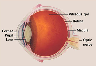
The optic nerve
The optic nerve is a bundle of more than 1 million nerve fibers. It connects the retina to the brain. (See diagram above.) The retina is the light-sensitive tissue at the back of the eye. A healthy optic nerve is necessary for good vision.

How does the optic nerve get damaged by open-angle glaucoma?

Several large studies have shown that eye pressure is a major risk factor for optic nerve damage. In the front of the eye is a space called the anterior chamber. A clear fluid flows continuously in and out of the chamber and nourishes nearby tissues. The fluid leaves the chamber at the open angle where the cornea and iris meet. (See diagram below.) When the fluid reaches the angle, it flows through a spongy meshwork, like a drain, and leaves the eye.
In open-angle glaucoma, even though the drainage angle is “open”, the fluid passes too slowly through the meshwork drain. Since the fluid builds up, the pressure inside the eye rises to a level that may damage the optic nerve. When the optic nerve is damaged from increased pressure, open-angle glaucoma-and vision loss—may result. That’s why controlling pressure inside the eye is important.
Another risk factor for optic nerve damage relates to blood pressure. Thus, it is important to also make sure that your blood pressure is at a proper level for your body by working with your medical doctor.
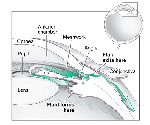
Fluid Pathway
Fluid pathway is shown in teal.

Can I develop glaucoma if I have increased eye pressure?

Not necessarily. Not every person with increased eye pressure will develop glaucoma. Some people can tolerate higher levels of eye pressure better than others. Also, a certain level of eye pressure may be high for one person but normal for another.
Whether you develop glaucoma depends on the level of pressure your optic nerve can tolerate without being damaged. This level is different for each person. That’s why a comprehensive dilated eye exam is very important. It can help your eye care professional determine what level of eye pressure is normal for you.

Can I develop glaucoma without an increase in my eye pressure?

Yes. Glaucoma can develop without increased eye pressure. This form of glaucoma is called low-tension or normal-tension glaucoma. It is a type of open-angle glaucoma.

Who is at risk for open-angle glaucoma?

Anyone can develop glaucoma. Some people, listed below, are at higher risk than others:
  • African Americans over age 40
  • Everyone over age 60, especially Mexican Americans
  • People with a family history of glaucoma
A comprehensive dilated eye exam can reveal more risk factors, such as high eye pressure, thinness of the cornea, and abnormal optic nerve anatomy. In some people with certain combinations of these high-risk factors, medicines in the form of eyedrops reduce the risk of developing glaucoma by about half.

Glaucoma Symptoms

At first, open-angle glaucoma has no symptoms. It causes no pain. Vision stays normal. Glaucoma can develop in one or both eyes.
Without treatment, people with glaucoma will slowly lose their peripheral (side) vision. As glaucoma remains untreated, people may miss objects to the side and out of the corner of their eye. They seem to be looking through a tunnel. Over time, straight-ahead (central) vision may decrease until no vision remains.
Normal Vision
Normal Vision.
Glaucoma Vision
The same scene as viewed by a person with glaucoma.
Glaucoma Detected

How is glaucoma detected?

Glaucoma is detected through a comprehensive dilated eye exam that includes the following:
Visual acuity test. This eye chart test measures how well you see at various distances.
Visual field test. This test measures your peripheral (side vision). It helps your eye care professional tell if you have lost peripheral vision, a sign of glaucoma.
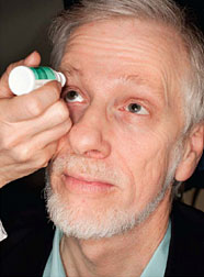
Dilated eye exam. In this exam, drops are placed in your eyes to widen, or dilate, the pupils. Your eye care professional uses a special magnifying lens to examine your retina and optic nerve for signs of damage and other eye problems. After the exam, your close-up vision may remain blurred for several hours.
Tonometry is the measurement of pressure inside the eye by using an instrument called a tonometer. Numbing drops may be applied to your eye for this test. A tonometer measures pressure inside the eye to detect glaucoma.
Pachymetry is the measurement of the thickness of your cornea. Your eye care professional applies a numbing drop to your eye and uses an ultrasonic wave instrument to measure the thickness of your cornea.

Can glaucoma be cured?

No. There is no cure for glaucoma. Vision lost from the disease cannot be restored.

Glaucoma Treatments

Immediate treatment for early-stage, open-angle glaucoma can delay progression of the disease. That’s why early diagnosis is very important.
Glaucoma treatments include medicines, laser trabeculoplasty, conventional surgery, or a combination of any of these. While these treatments may save remaining vision, they do not improve sight already lost from glaucoma.
Medicines. Medicines, in the form of eyedrops or pills, are the most common early treatment for glaucoma. Taken regularly, these eyedrops lower eye pressure. Some medicines cause the eye to make less fluid. Others lower pressure by helping fluid drain from the eye.
Before you begin glaucoma treatment, tell your eye care professional about other medicines and supplements that you are taking. Sometimes the drops can interfere with the way other medicines work.
Glaucoma medicines need to be taken regularly as directed by your eye care professional. Most people have no problems. However, some medicines can cause headaches or other side effects. For example, drops may cause stinging, burning, and redness in the eyes.
Many medicines are available to treat glaucoma. If you have problems with one medicine, tell your eye care professional. Treatment with a different dose or a new medicine may be possible.
Because glaucoma often has no symptoms, people may be tempted to stop taking, or may forget to take, their medicine. You need to use the drops or pills as long as they help control your eye pressure. Regular use is very important.
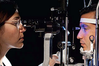
Tonometer that measures pressure.
A tonometer measures pressure inside the eye to detect glaucoma.
Make sure your eye care professional shows you how to put the drops into your eye. For tips on using your glaucoma eyedrops, see the inside back cover of this booklet.
Laser trabeculoplasty. Laser trabeculoplasty helps fluid drain out of the eye. Your doctor may suggest this step at any time. In many cases, you will need to keep taking glaucoma medicines after this procedure.
Laser trabeculoplasty is performed in your doctor’s office or eye clinic. Before the surgery, numbing drops are applied to your eye. As you sit facing the laser machine, your doctor holds a special lens to your eye. A high-intensity beam of light is aimed through the lens and reflected onto the meshwork inside your eye. You may see flashes of bright green or red light. The laser makes several evenly spaced burns that stretch the drainage holes in the meshwork. This allows the fluid to drain better.
Like any surgery, laser surgery can cause side effects, such as inflammation. Your doctor may give you some drops to take home for any soreness or inflammation inside the eye. You will need to make several follow-up visits to have your eye pressure and eye monitored.
If you have glaucoma in both eyes, usually only one eye will be treated at a time. Laser treatments for each eye will be scheduled several days to several weeks apart.
Studies show that laser surgery can be very good at reducing the pressure in some patients. However, its effects can wear off over time. Your doctor may suggest further treatment.
Conventional surgery. Conventional surgery makes a new opening for the fluid to leave the eye. (See diagram on the next page.) Your doctor may suggest this treatment at any time. Conventional surgery often is done after medicines and laser surgery have failed to control pressure.
Conventional surgery, called trabeculectomy, is performed in an operating room. Before the surgery, you are given medicine to help you relax. Your doctor makes small injections around the eye to numb it. A small piece of tissue is removed to create a new channel for the fluid to drain from the eye. This fluid will drain between the eye tissue layers and create a blister-like “filtration bleb.”
For several weeks after the surgery, you must put drops in the eye to fight infection and inflammation. These drops will be different from those you may have been using before surgery.
Conventional surgery is performed on one eye at a time. Usually the operations are four to six weeks apart.
Conventional surgery is about 60 to 80 percent effective at lowering eye pressure. If the new drainage opening narrows, a second operation may be needed. Conventional surgery works best if you have not had previous eye surgery, such as a cataract operation.
Sometimes after conventional surgery, your vision may not be as good as it was before conventional surgery. Conventional surgery can cause side effects, including cataract, problems with the cornea, inflammation, infection inside the eye, or low eye pressure problems. If you have any of these problems, tell your doctor so a treatment plan can be developed.

What are some other forms of glaucoma and how are they treated?

Open-angle glaucoma is the most common form. Some people have other types of the disease.
In low-tension or normal-tension glaucoma, optic nerve damage and narrowed side vision occur in people with normal eye pressure. Lowering eye pressure at least 30 percent through medicines slows the disease in some people. Glaucoma may worsen in others despite low pressures.
A comprehensive medical history is important to identify other potential risk factors, such as low blood pressure, that contribute to low-tension glaucoma. If no risk factors are identified, the treatment options for low-tension glaucoma are the same as for open-angle glaucoma.
In angle-closure glaucoma, the fluid at the front of the eye cannot drain through the angle and leave the eye. The angle gets blocked by part of the iris. People with this type of glaucoma may have a sudden increase in eye pressure. Symptoms include severe pain and nausea, as well as redness of the eye and blurred vision. If you have these symptoms, you need to seek treatment immediately. This is a medical emergency. If your doctor is unavailable, go to the nearest hospital or clinic. Without treatment to restore the flow of fluid, the eye can become blind. Usually, prompt laser surgery and medicines can clear the blockage, lower eye pressure, and protect vision.
In congenital glaucoma, children are born with a defect in the angle of the eye that slows the normal drainage of fluid. These children usually have obvious symptoms, such as cloudy eyes, sensitivity to light, and excessive tearing. Conventional surgery typically is the suggested treatment, because medicines are not effective and can cause more serious side effects in infants and be difficult to administer. Surgery is safe and effective. If surgery is done promptly, these children usually have an excellent chance of having good vision.
Conventional surgery.
Conventional surgery makes a new opening for the fluid to leave the eye.
Secondary glaucomas can develop as complications of other medical conditions. For example, a severe form of glaucoma is called neovascular glaucoma, and can be a result from poorly controlled diabetes or high blood pressure. Other types of glaucoma sometimes occur with cataract, certain eye tumors, or when the eye is inflamed or irritated by a condition called uveitis. Sometimes glaucoma develops after other eye surgeries or serious eye injuries. Steroid drugs used to treat eye inflammations and other diseases can trigger glaucoma in some people. There are two eye conditions known to cause secondary forms of glaucoma.
Pigmentary glaucoma occurs when pigment from the iris sheds off and blocks the meshwork, slowing fluid drainage.
Pseudoexfoliation glaucoma occurs when extra material is produced and shed off internal eye structures and blocks the meshwork, again slowing fluid drainage.
Depending on the cause of these secondary glaucomas, treatment includes medicines, laser surgery, or conventional or other glaucoma surgery.

What research is being done?

Through studies in the laboratory and with patients, NEI is seeking better ways to detect, treat, and prevent vision loss in people with glaucoma. For example, researchers have discovered genes that could help explain how glaucoma damages the eye.
NEI also is supporting studies to learn more about who is likely to get glaucoma, when to treat people who have increased eye pressure, and which treatment to use first.

What You Can Do

If you are being treated for glaucoma, be sure to take your glaucoma medicine every day. See your eye care professional regularly.
You also can help protect the vision of family members and friends who may be at high risk for glaucoma-African Americans over age 40; everyone over age 60, especially Mexican Americans; and people with a family history of the disease. Encourage them to have a comprehensive dilated eye exam at least once every two years. Remember that lowering eye pressure in the early stages of glaucoma slows progression of the disease and helps save vision.
Medicare covers an annual comprehensive dilated eye exam for some people at high risk for glaucoma. These people include those with diabetes, those with a family history of glaucoma, and African Americans age 50 and older.

What should I ask my eye care professional?

You can protect yourself against vision loss by working in partnership with your eye care professional. Ask questions and get the information you need to take care of yourself and your family.

What are some questions to ask?

About my eye disease or disorder…
  • What is my diagnosis?
  • What caused my condition?
  • Can my condition be treated?
  • How will this condition affect my vision now and in the future?
  • Should I watch for any particular symptoms and notify you if they occur?
  • Should I make any lifestyle changes?
About my treatment…
  • What is the treatment for my condition?
  • When will the treatment start and how long will it last?
  • What are the benefits of this treatment and how successful is it?
  • What are the risks and side effects associated with this treatment?
  • Are there foods, medicines, or activities I should avoid while I’m on this treatment?
  • If my treatment includes taking medicine, what should I do if I miss a dose?
  • Are other treatments available?
About my tests…
  • What kinds of tests will I have?
  • What can I expect to find out from these tests?
  • When will I know the results?
  • Do I have to do anything special to prepare for any of the tests?
  • Do these tests have any side effects or risks?
  • Will I need more tests later?
Other suggestions
  • If you don’t understand your eye care professional’s responses, ask questions until you do understand.
  • Take notes or get a friend or family member to take notes for you. Or, bring a tape recorder to help you remember the discussion.
  • Ask your eye care professional to write down his or her instructions to you.
  • Ask your eye care professional for printed material about your condition.
  • If you still have trouble understanding your eye care professional’s answers, ask where you can go for more information.
  • Other members of your healthcare team, such as nurses and pharmacists, can be good sources of information. Talk to them, too.
Today, patients take an active role in their health care. Be an active patient about your eye care.

Loss of Vision

If you have lost some sight from glaucoma, ask your eye care professional about low vision services and devices that may help you make the most of your remaining vision. Ask for a referral to a specialist in low vision. Many community organizations and agencies offer information about low vision counseling, training, and other special services for people with visual impairments.

How should I use my glaucoma eyedrops?

If eyedrops have been prescribed for treating your glaucoma, you need to use them properly, as instructed by your eye care professional. Proper use of your glaucoma medication can improve the medicine’s effectiveness and reduce your risk of side effects.
To properly apply your eyedrops, follow these steps:
  • Wash your hands.
  • Hold the bottle upside down.
  • Tilt your head back.
  • Hold the bottle in one hand and place it as close as possible to the eye.
  • With the other hand, pull down your lower eyelid. This forms a pocket.
  • Place the prescribed number of drops into the lower eyelid pocket. If you are using more than one eyedrop, be sure to wait at least 5 minutes before applying the second eyedrop.
  • Close your eye OR press the lower lid lightly with your finger for at least 1 minute. Either of these steps keeps the drops in the eye and helps prevent the drops from draining into the tear duct, which can increase your risk of side effects.

Iron-Rich Foods By Mary Anne Dunkin


Spinach may not give you superhuman strength to fight off villains like Popeye's nemesis Bluto, but this leafy green and other foods containing iron can help you fight a different type of enemy -- iron-deficiency anemia.
Iron-deficiency anemia, the most common form of anemia, is a decrease in the number of red blood cells caused by too little iron. Without sufficient iron, your body can't produce enough hemoglobin, a substance in red blood cells that makes it possible for them to carry oxygen to the body's tissues. As a result, you may feel weak, tired, and irritable.
spinach and steak
About 20% of women, 50% of pregnant women, and 3% of men do not have enough iron in their body. The solution, in many cases, is to consume more foods high in iron.

How Your Body Uses Iron in Food

When you eat food with iron, iron is absorbed into your body mainly through the upper part of your small intestine.
There are two forms of dietary iron: heme and nonheme. Heme iron is derived from hemoglobin. It is found in animal foods that originally contained hemoglobin, such as red meats, fish, and poultry. Your body absorbs the most iron from heme sources. Nonheme iron is from plant sources.

Iron-Rich Foods

Very good sources of heme iron, with 3.5 milligrams or more per serving, include:
  • 3 ounces of beef or chicken liver
  • 3 ounces of clams, mollusks, or mussels
  • 3 ounces of oysters
Good sources of heme iron, with 2.1 milligrams or more per serving, include:
  • 3 ounces of cooked beef
  • 3 ounces of canned sardines, canned in oil
  • 3 ounces of cooked turkey
Other sources of heme iron, with 0.7 milligrams or more per serving, include:
  • 3 ounces of chicken
  • 3 ounces of halibut, haddock, perch, salmon, or tuna
  • 3 ounces of ham
  • 3 ounces of veal
Iron in plant foods such as lentils, beans, and spinach is nonheme iron. This is the form of iron added to iron-enriched and iron-fortified foods. Our bodies are less efficient at absorbing nonheme iron, but most dietary iron is nonheme iron.